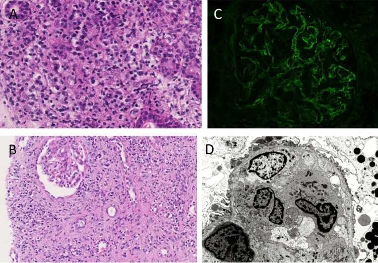
Figure 2.
Light microscopy, immunofluorescence and electron microscopy findings. (A) Light microscopy of the right submandibular gland shows massive lymphoplasmacytic infiltration with fibrosis (H&E; original magnification, ×200). (B) Light microscopy of the right kidney shows diffuse lymphoplasmacytic infiltration, with marked interstitial fibrosis (H&E; original magnification, ×100). Angitis, granulomatous lesions, neutrophil infiltration and advanced tubulitis were not found. (C) Immunofluorescence of the glomerulus showing diffuse granular staining for IgG in the glomerular basement membrane. (D) Electron microscopy of the glomerulus showing focal effacement of podocyte foot processes, with scattered subepithelial electron-dense deposits.

