
Figure 1.
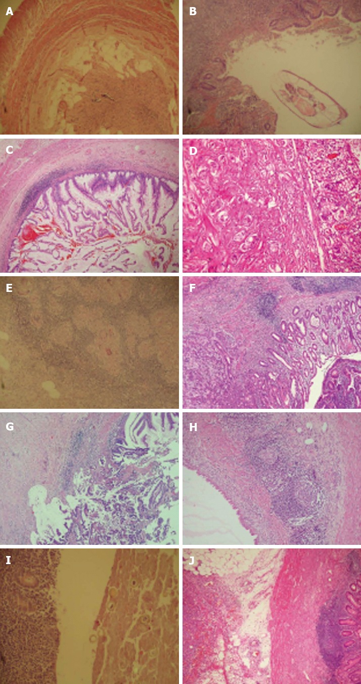
Unusual histopathologic findings. A: Appendix vermiformis showing fibrous obliteration [hematoxylin and eosin (HE) × 40]; B: View of the enterobius vermiformis in the lumen of appendix vermiformis (HE × 100); C: Mucinous cystadenoma showing proliferation of neoplastic adenomatous epithelium, which exhibits low-grade dysplasia (HE × 100); D: Carcinoid tumor of the appendix showing rounded nests and tubules of tumor cells with uniform nuclei (HE × 200); E: Granulomatous inflammation. Submucosal granuloma with central necrosis (HE × 40); F: Moderately differentiated adenocarcinoma showing infiltration of the mucosa and submucosa of the appendiceal wall (HE × 100); G: Adenocarcinoma of the appendix showing associated mucocele on the top right side (HE × 100); H: Mucocele showing a unilocular dilated appendiceal wall lined with flattened epithelial cells (HE × 100); I: Eggs of Taenia sup are present in the lumen of appendix vermiformis (HE × 100); J: Serosa of the appendiceal wall showing diffuse large B cell lymphoma infiltration (HE × 40).