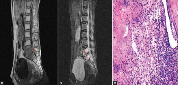
Figure 1.

Classical paradiscal tuberculosis of the spine (a) T1W MRI sagittal view showing hypointense L5 and S1 vertebral bodies and arrow shows elevation of posterior longitudinal ligament (b) T2W MRI sagittal view showing hyperintense lesions involving L5 and S1 vertebral bodies suggestive of edema/inflammation (c) Spine TB-10 × H and E, showing caseous necrosis, epithelioid cell, giant cell and bone fragment
