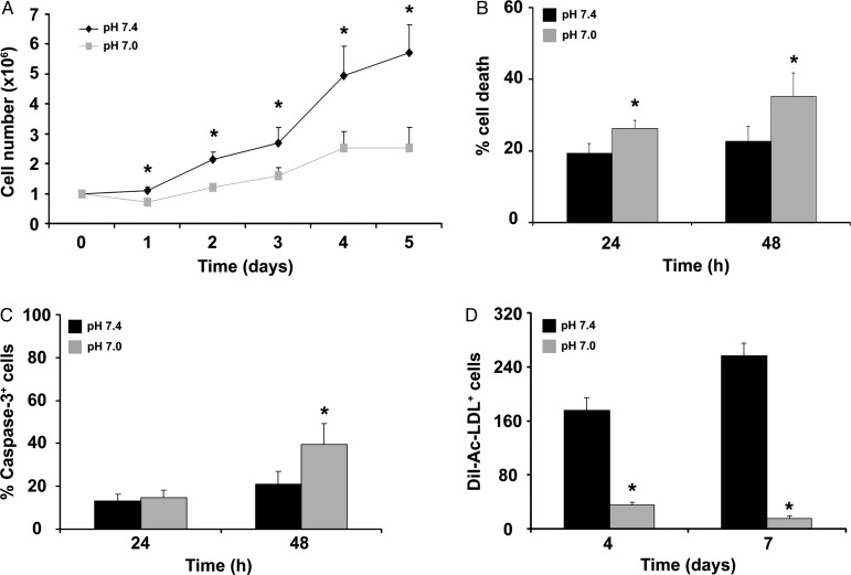
Figure 1.
Acidosis effect on ckit+ cell proliferation, death, and endothelial differentiation. (A) Acidosis inhibited the progressive increase in ckit+ cell number at pH 7.4 (n = 5). (B) Acidosis enhanced cell death, as assessed by FACS analysis of PI-stained cells (n = 9). (C) FACS analysis for caspase-3 (n = 3). (D) At 4 and 7 days, the number of DiI-Ac-LDL+ cells was lower at pH 7.0 vs. 7.4 (n = 3 in duplicate). Statistical significance: *P < 0.05 for pH 7.4 vs. 7.0.

