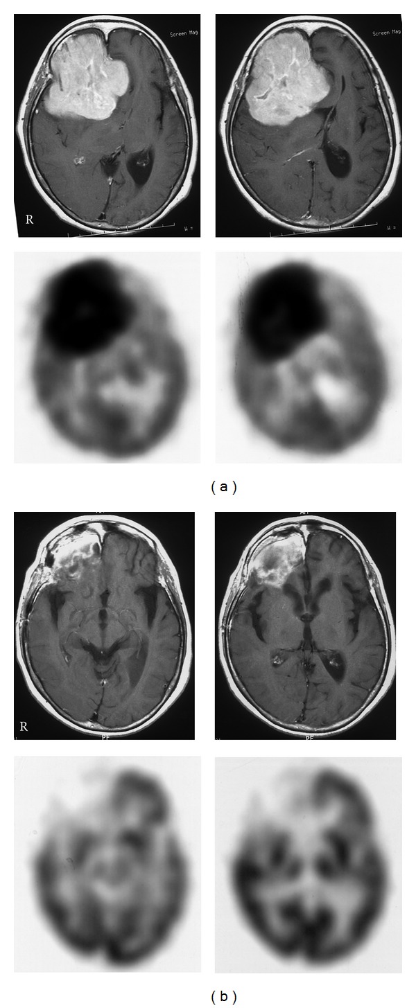
Figure 5.

Contrast-enhanced T1-weighted MR (upper) and 18F-FDG PET (lower) images in a patient with PCNSL in the right frontal lobe before (a) and after (b) the first cycle of chemotherapy. MR images show a well-enhanced large mass lesion in the right frontal lobe, and PET images show a huge 18F-FDG uptake in the lesion before treatment (a). After the first chemotherapy, MR images show a residual enhanced lesion in the right frontal lobe; however, PET images show no increased 18F-FDG uptake in the lesion (b).
