Figure 5.

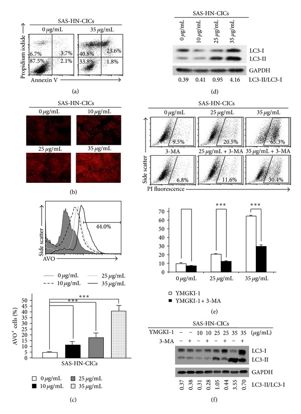
YMGKI-1 treatment induces autophagic cell death in HN-CICs. (a) SAS-HN-CICs treated with 35 μg/mL of YMGKI-1 were costained with Annexin V and Propidium iodide (PI) and examined by flow cytometry. (b) SAS-HN-CICs were treated with YMGKI-1 at different concentration for 24 hr, afterward, stained with acridine orange, and observed under a red filter fluorescence microscope. (c) Acridine orange (AO) positively stained cells of SAS-HN-CICs in (b) were quantitated by flow cytometric analysis (upper panel). The mean fluorescence intensity of AO stained cells was calculated (lower panel). Data are means ± SD of triplicate samples from three experiments (***P < 0.005). (d) Crude cell extract proteins of YMGKI-1-treated SAS-HN-CICs were collected, electrophorized, and further analyzed by immunoblotting against anti-LC3 or anti-GAPDH serum as indicated. (e) SAS-HN-CICs cells were either singly treated with 0, 25, or 35 μg/mL of YMGKI-1, or cotreated with 3-Methylamphetamine (3-MA), an autophagy inhibitor, for 24 hr, afterward, stained with propidium iodide (PI), and then examined by flow cytometry. Data are means ± SD of triplicate samples from three experiments (***P < 0.005). (f) Crude cell extract proteins of SAS-HN-CICs singly treated with YMGKI-1 or cotreated with 3-MA were isolated and analyzed by immunoblotting against anti-LC3 or anti-GAPDH serum as indicated. The immunoactive signal of GAPDH protein of different crude cell extracts was referred as loading control. The same concentration (0.03%) of vehicle (DMSO) was added to all control groups.
