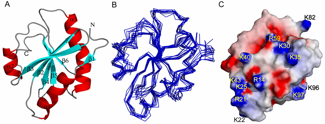
Figure 2.
Solution structure of TgADF. (A) Ribbon diagram of lowest energy structure of TgADF showing six stranded mixed β-sheet surrounded by three α-helices and a C-terminal helical turn. The individual β strands and α helices are labeled. (B) Superimposition of backbone traces from final ensemble of 10 structures with lowest target function. (C) Electrostatic potential of TgADF generated with GRASP. The positively and negatively charged groups are shown in blue and red, respectively.

