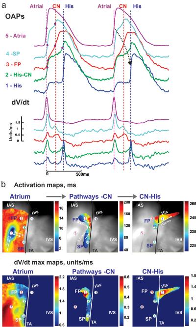
Fig. 3.
a Optical APs and their maximum derivatives from five recording sites in the human AVN preparation during atrial pacing at cycle length 1000ms (recording sites are solid circles in b and c, superimposed on the optical field of view, which is 30×30 mm2) b Separate activation maps of the right atrium, AVN and His bundle allow tracking of conduction with conduction velocities. Black line represents the tricuspid valve annulus c dV/dtmax maps in the same areas. FP: fast pathway, SL: slow pathway, IAS: inter-atrial septum, IVS: inter-ventricular septum, CN: compact node (Reproduced from Fedorov et al. [25] with permission)

