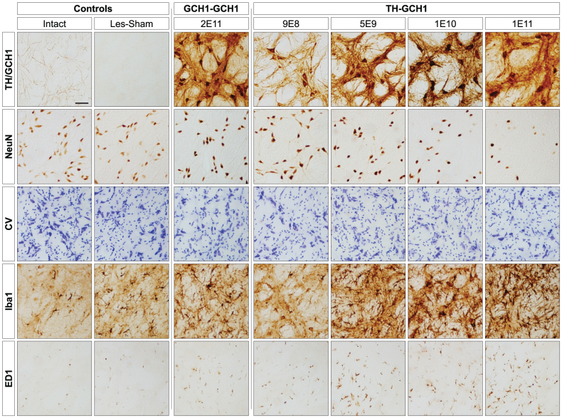
Figure 5. High magnification images of neurons in the globus pallidus (GP).
Transgene immunoreactivity (row 1, TH staining depicted in all panels except GCH1-GCH1 which is a GCH1 staining) was present in GP in all vector-treated animals filling cell bodies, dendritic processes and axon terminals. Loss of NeuN positive cells in GP was seen in animals injected with 1E11 gc of the TH-GCH1 vector (row 2, far right) but also to a lesser extent in the 1E10 group. Nissl stained specimens from adjacent sections confirmed loss of neurons (row 3). In the same region, there was an increase in the microglial marker Iba1 (row 4) seen as a dose-dependent increase in arborized reactive cells. ED1 (equivalent to human CD68) immunoreactivity indicated presence of macrophages. Scale bar in top left panel represent 0.2 mm in all panels.

