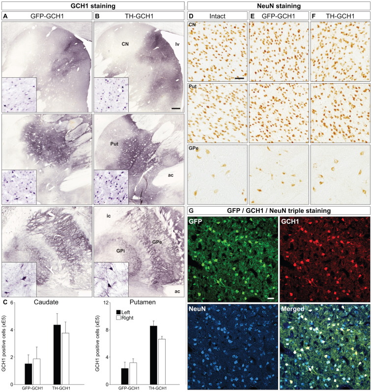
Figure 7. Transgene expression in MPTP-treated monkeys.
Transgene expression from both AAV vectors encoding GFP-GCH1 (A) and TH-GCH1 (B) were visualized with the GCH1 antibody, which showed GCH1 immunoreactivity in the putamen (Put, row 1), caudate nucleus (CN, row 2) and globus pallidus (GP, row 3). Interestingly, both the external and internal segment of GP had GCH1 immunoreactive cells and projections (insets). Stereological quantification of GCH1 positive neurons was performed in both putamen and caudate nucleus bilaterally (C). NeuN stainings were performed in adjacent sections and are shown from Put, CN and GPe (E-F). Co-expression of GFP and GCH1 in the transduced neurons was confirmed with confocal microscopy (G). Whereas the GCH1 staining was robust, the expression of the TH transgene could not be confirmed in the TH-GCH1 injected monkeys. ac = anterior commissure, CN = caudate nucleus, GPi/GPe = globus pallidus internal/external segment, lv = lateral ventricle, Put = putamen. Scale bar in (B) represent 0.5 mm and (D) and (G) represents 50 μm. GCH1 insets in (A–B) have the same magnification as (D). Cell counts in (C) represent mean SEM.

