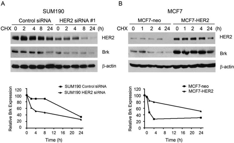
Fig. 2.
Role of HER2 in Brk protein stability. (A) SUM190 cells were transfected with a HER2-specific siRNA#1 or control siRNA for 48 h. The cells were then exposed to 10 μM cycloheximide (CHX) in 0.5% FBS medium for the indicated times. The levels of HER2 and Brk were detected by Western blotting with the indicated antibodies. β-actin was used as a loading control. Levels of HER2 and Brk relative to the internal control β-actin were quantified and plotted as shown. (B) MCF7-neo and MCF7-HER2 cells were exposed to CHX (10 μM) in 0.5% FBS medium for the indicated times. The levels of HER2 and Brk were detected by Western blotting with the indicated antibodies. β-actin was used as a loading control. Levels of HER2 and Brk relative to the internal control β-actin were quantified and plotted as shown.

