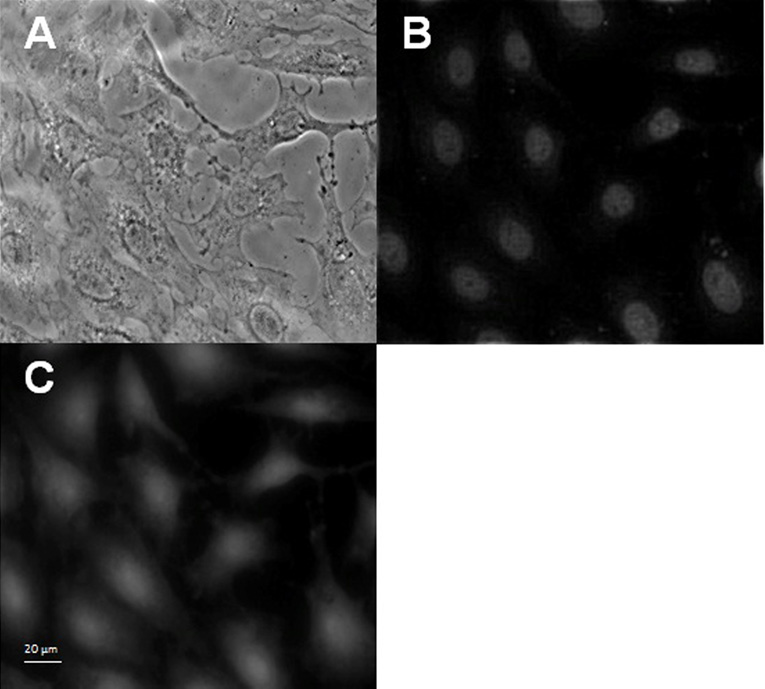
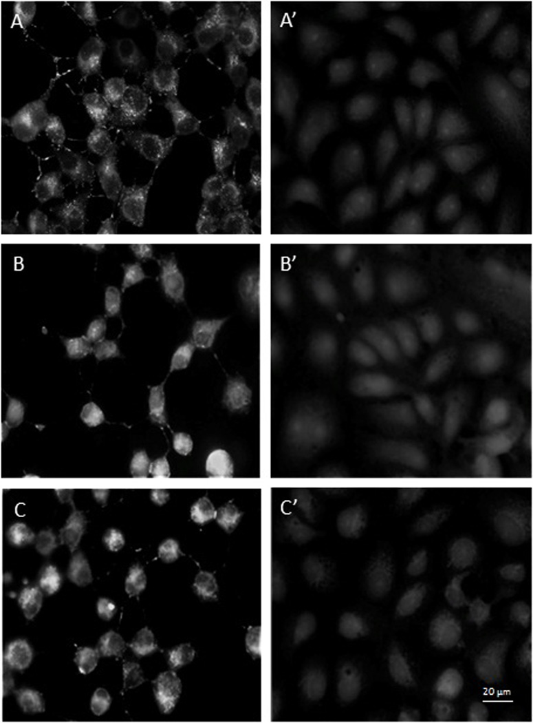
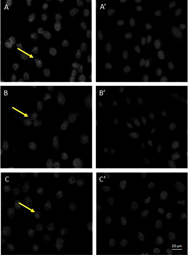
Figure 5.
Fluorescence microscopic observations of control Vero cells and p-EGCG treated HSV-1 infection of Vero Cells. 5.1. Control Vero cells monolayer (400×). (A) Phase contrast; (B) DAPI stain; (C) GFP. 5.2. GFP expression and 5.3. DAPI stain at 8–12 hours post-infection for HSV-1 (+/− p-EGCG) of Vero cells at (A) 8 hours no p-EGCG (A’) 8 hours + p-EGCG; (B) 10 hours no p-EGCG; (B’) 10 hours + p-EGCG; (C) 12 hours no p-EGCG; (C’) 12 hours + p-EGCG. The arrows show the granulation and loss of margins in the HSV-1 infected cells.


