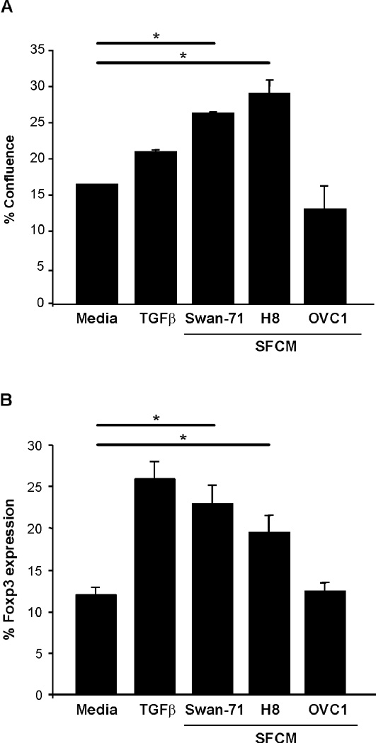
Figure 2. Trophoblast cells contribute to the differentiation of iTregs from maternal naïve T cells.
(A) In vitro differentiation of iTregs was performed in the presence of LSCM from Swan-71 and H8. As positive controls we performed the differentiation in the presence of recombinant TGFβ, and in the presence of media (just with IL-2) or LSCM from OVC1 as negative controls. LSCM from Swan and H8 cells induce a significant increase of Foxp3 expression evaluated by FACS analysis (*p<0.05, Student T-test); LSCM=CM. (B) Cellular proliferation was measured with an IncuCyte™ Long-term live cell imaging system and results are expressed as the percentage of confluence based in the image metrics data.

