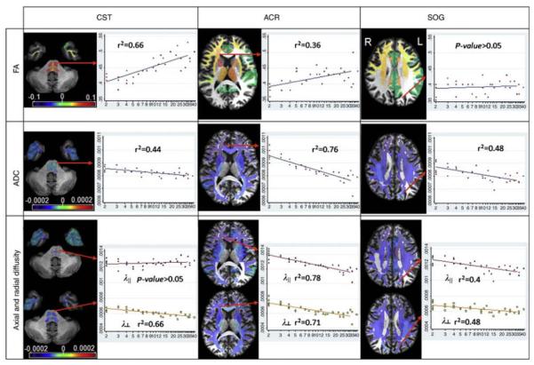
Fig. 5.
Actual fitting results for the fractional anisotropy (FA) (first row), apparent diffusion coefficient (ADC) (second row, in mm2/s), and axial and radial diffusivity (third row, in mm2/s), by age (in years, logarithmic scale), at representative locations. In the corticospinal tract (CST, first column), the FA increase can be explained by the radial diiffusivity decrease. In the anterior corona radiata (ACR, second column), the age-related changes in the axial diffusivity cause a weaker time-dependent FA change. In the white matter of the superior occipital gyrus (SOG, third column), the parallel decreases in both axial and radial diffusivity lead to no significant changes in FA. The orientation of the slices follows the radiologic convention (L left, R right) (printed with permission [72])

