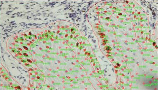
Figure 2.

Photomicrograph showing Image Pro Express used for counting of Ki-67 positivecells in basal, parabasal, and suprabasal layers of epithelium

Photomicrograph showing Image Pro Express used for counting of Ki-67 positivecells in basal, parabasal, and suprabasal layers of epithelium