Figure 1.

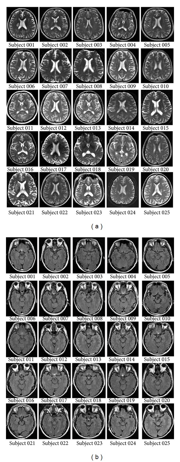
(a) Lesion locations of stroke patients on axial T2-weighted images acquired at the stroke onset when stroke patients were first enrolled in our hospital. (b) The morphology and signals of the brainstem of every stroke patient.

(a) Lesion locations of stroke patients on axial T2-weighted images acquired at the stroke onset when stroke patients were first enrolled in our hospital. (b) The morphology and signals of the brainstem of every stroke patient.