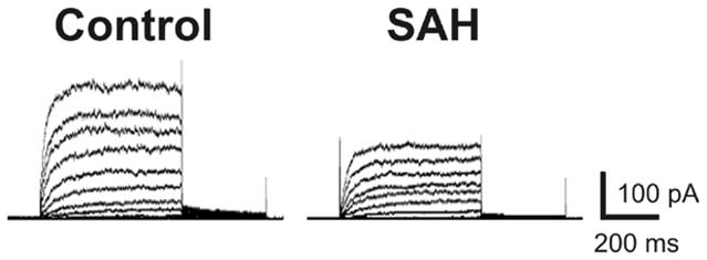
Figure 3. Voltage-dependent K+ channel currents are decreased in parenchymal arteriolar myocytes following SAH.

Representative traces of voltage-dependent K+ channel currents recorded using the conventional whole-cell patch clamp technique from control (cell capacitance: 8.3 pF) and SAH animals (cell capacitance: 8.2 pF).
