Fig. 1.
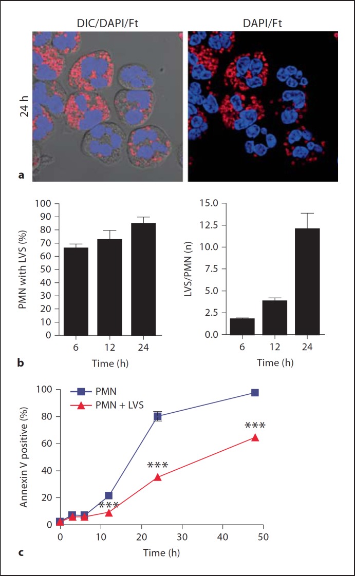
F. tularensis LVS infects human neutrophils and impairs their spontaneous apoptosis. Neutrophils from four independent donors were infected on different days with LVS for 3-48 h at 37°C in serum-free media. a Representative confocal images of infected PMNs at 24 h. Bacteria are shown in red and PMNs were detected using differential interference contrast optics and DAPI-staining of nuclear DNA (blue). b Quantitation of infection efficiency. Graphs show the percentage of infected PMNs and bacterial load per infected cell. Pooled data are the mean ± SEM (n = 4). c Apoptosis was assessed at the indicated time points using Annexin V-FITC staining and flow cytometry. Pooled data are the mean ± SEM (n = 4). *** p < 0.001 for control versus LVS-infected PMNs.

