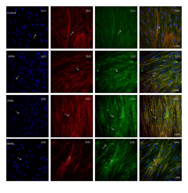
Figure 7.

Representative confocal laser microscopy images of hABMSCs cultured for 7 days in static conditions (a1–d1) or ELF-PEMFs induction (intensity, 6 Gauss) at 10 Hz/day (a2–d2), 50 Hz/day (a3–d3), and 100 Hz/day (a4–d4) groups; cell nuclei (a1–d4), actin filaments (b1–b4), vinculin (c1–c4), and merged images (d1–d4) of the fluorescence stains. Confocal laser microscopy images showed more intense observation at ELF-PEMFs induction groups compared to control group (arrows: cell direction).
