
Figure 1.
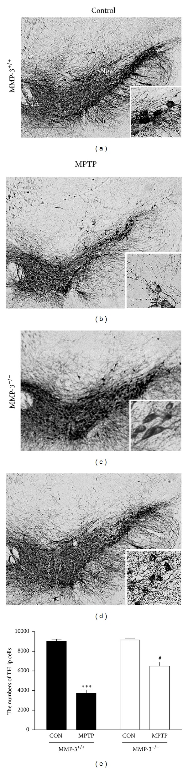
MPTP-induced neurotoxicity is attenuated in the SNpc of MMP-3−/− mouse brain. (a) Animals (MMP-3+/+ or MMP-3−/− mice) receiving PBS as a control (a and c) and MPTP (b and d) were sacrificed 7 days after the last MPTP injection. Insets, higher magnifications of (a–c). The brain tissues were cut into 30 μm thick coronal sections using a sliding microtome and immunostained with an antibody against the DA neuronal marker TH. Scale bar, 300 μm. (e) Bars represent the number of TH-ip neurons in the SN after indicated treatment in the absence (MMP-3−/−) or presence (MMP-3+/+) of MMP-3. Five to six animals were used for each experimental group. Two-way ANOVA with Bonferroni post hoc test (F(1,15) = 17.57, P < 0.001), ***P < 0.001, significantly different from PBS-injected MMP-3+/+ mice; # P < 0.05, significantly different from MPTP-injected MMP-3+/+ mice. SNpc, substantia nigra pars compacta; VTA, ventral tegmental area; SNr, substantia nigra reticulata.