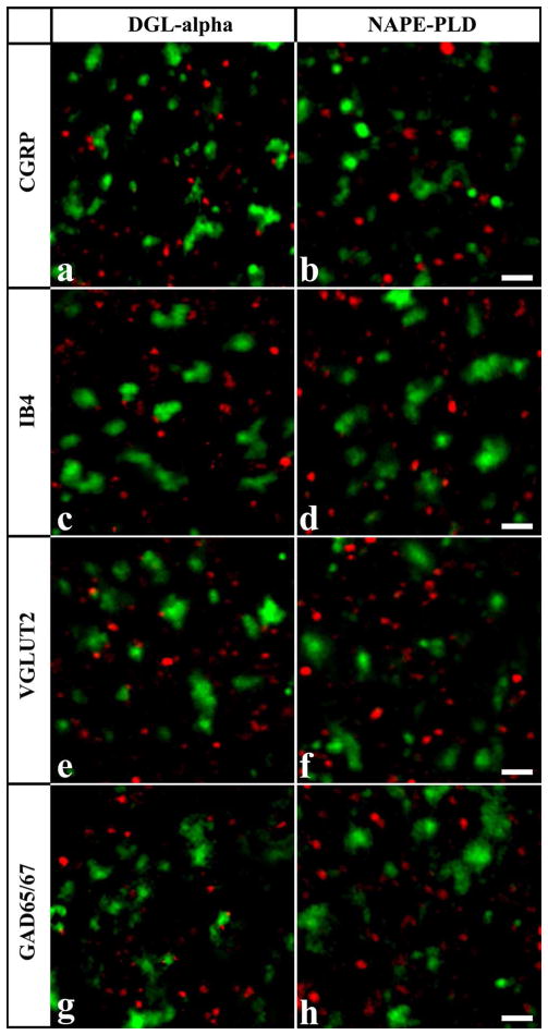
Figure 2.
Co-localization of DGLα and NAPE-PLD with axonal markers. Micrographs of single 1μm thick laser scanning confocal optical sections assessing co-localization between immunolabeling for DGLα (red; a, c, e, g) or NAPE-PLD (red; b, d, f, h) and immunoreactivity for markers that are specific for axon terminals of specific populations of peptidergic (CGRP, green; a, b) and non-peptidergic (IB4-binding, green; c, d) nociceptive primary afferents, as well as for axon terminals of putative excitatory (VGLUT2, green; e, f) and inhibitory (GAD65/67, green; g, h) intrinsic neurons in the superficial spinal dorsal horn. Note that mixed colors (yellow) that may indicate double labeled structures are not observed on the superimposed images. Scale bar: 2 μm.

