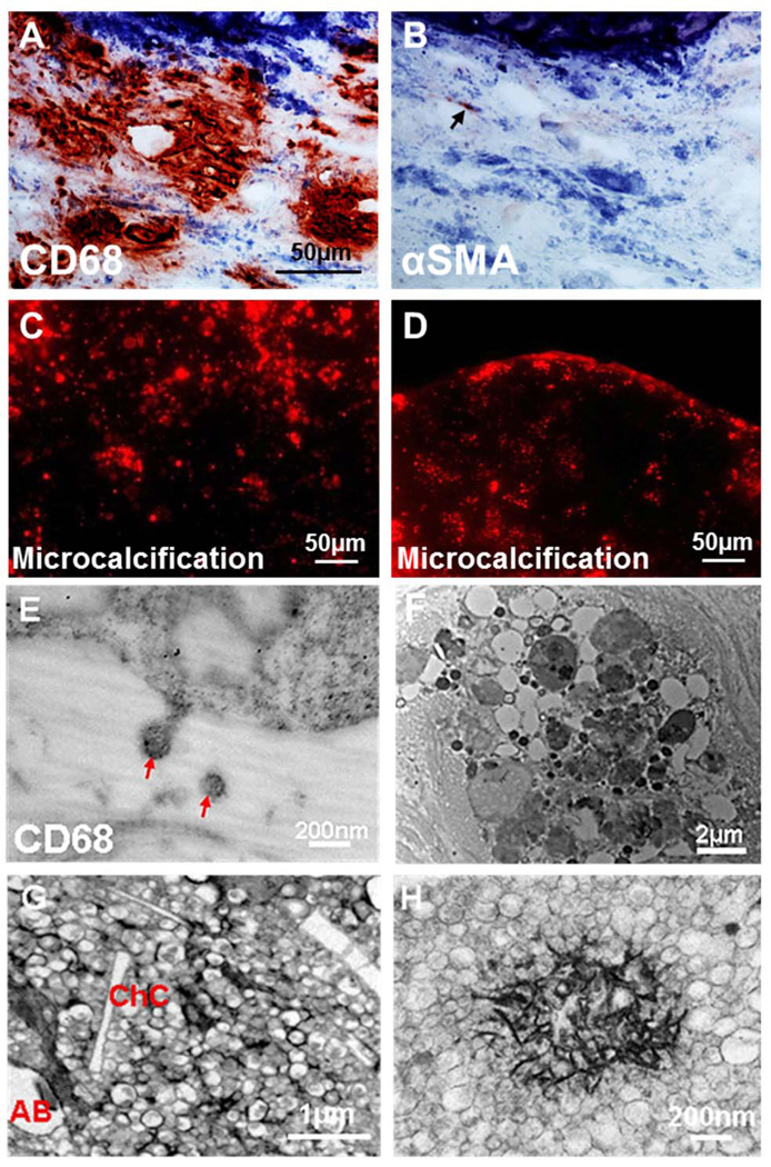
Figure 1. Macrophage-derived MVs contribute to atherosclerotic microcalcification.
A, Calcified vesicular structures (hematoxylin; blue) adjacent to macrophages (CD68; red) in human plaques (n=127). B, Few to no αSMA-positive SMCs were noted in the same area. C, Ex vivo hydroxyapatite-binding NIRF imaging agent detected vesicular microcalcification (red) within human and, D, apoE−/− mouse plaques. E–F, Ultrastructure of human calcified plaques: E, Macrophage releases CD68-positive MVs (immunogold). F, Vesicles of different sizes and appearance. G–H, Ultrastructure of calcified CRD apoE−/− mouse plaque: G, MVs near cholesterol crystals (ChC; AB apoptotic bodies). H, MV (30–300 nm) hydroxyapatite crystallization.

