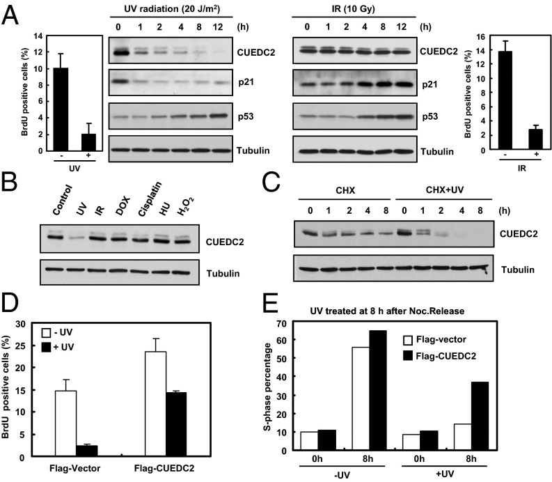
Fig. 1.
CUEDC2 is degraded during UV-induced G1 arrest and its overexpression overcomes such an arrest. (A) Immunoblot analysis of CUEDC2 and other proteins in U2OS cells treated with UV-C (20 J/m2) irradiation or IR (10 Gy). The proportion of BrdU-positive cells were analyzed by FACS. (B) Detection of CUEDC2 protein levels in response to various DNA damage agents in U2OS cells. (C) U2OS cells were pretreated for 30 min with cycloheximide (20 mM) followed by UV treatment. CUEDC2 protein levels were determined as indicated. (D) MCF-10A stably expressing Flag-Vector or Flag-CUEDC2 cells were treated with or without UV-C (20 J/m2) irradiation. After an additional 4 h, cells were pulsed with BrdU (10 μM) for 1 h. The proportion of BrdU-positive cells were analyzed by FACS (error bars indicate SD; n = 3). (E) HeLa cells were transfected with Flag-Vector or Flag-CUEDC2, and 24 h later, cells were synchronized at mitosis by thymidine–nocodazole treatment, and then treated with or without UV-C (20 J/m2) irradiation at 8 h after nocodazole release and harvested at indicated times. Flag-Vector or Flag-CUEDC2–positive cells were analyzed for cell cycle distribution by FACS. The percentage of S-phase cells is shown in the histogram.

