
Fig. 5.
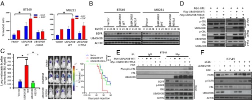
Oncogenic role of UBASH3B requires phosphatase activity targeting CBL phosphorylation and EGFR degradation (A) Transwell invasion of cells expressing WT or mutant UBASH3B (H391A) in the presence or absence of EGF as a chemoattractant. *P < 0.05, paired two-tailed t test. (B) EGFR and UBASH3B expression assessed by Western blot analysis. (C) (Left) BLI values of lung metastasis development in female NOD/SCID mice at 6 wk after injection with control and WT and mutant UBASH3B cells via the lateral tail vein. Data are mean ± SEM; n = 12 per category. *P < 0.05, unpaired two-sided Student t test. (Center) Representative BLI images of mice at 6 wk postinjection. (Right) Kaplan–Meier analysis. **P < 0.01, log-rank test. (D) Coimmunoprecipitation assays conducted on HEK293T with transient expression of CBL and WT or mutant UBASH3B in the presence or absence of EGF. (E) Coimmunoprecipitation assays conducted on BT549 stably expressing WT or mutant UBASH3B in the presence or absence of EGF. (F) Western blot analysis of EGFR, UBASH3B, and CBL protein expression in BT549 depleted with UBASH3B and/or CBL. Error bars represent mean ± SEM.