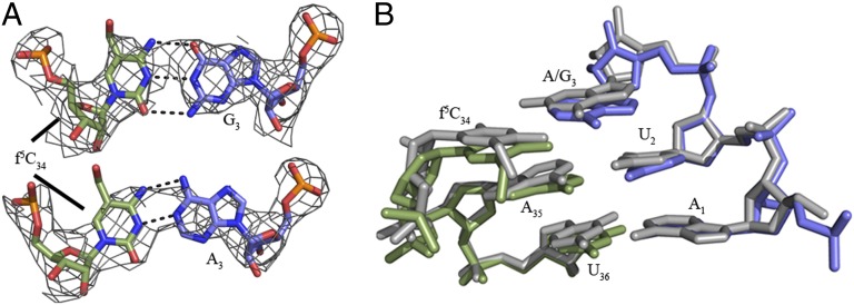
Fig. 3.
Geometry of the wobble base pair and codon•anticodon interaction. (A) The electron density shows that both the f5C34•G3 and f5C34•A3 base pairs are in Watson–Crick geometry. ASL carbons are colored green, and mRNA carbons are blue (m2FO-dFC contoured at 1.5 σ). (B) Superposition of the  AUG complex with the
  AUA structure aligned with respect to the mRNA residues. The overlay reveals a nearly identical orientation of the codon•anticodon interaction between the AUA-bound and AUG-bound structures. The ASL and mRNA residues for the AUA-bound structures are green and blue, respectively. The ASL and mRNA residues of the AUG-bound structure are both gray.

