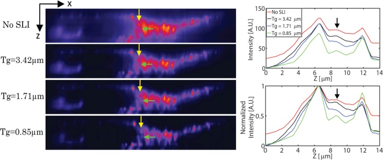
Fig. 5.
xz sections of the fine glomeruli and convoluted tubules structure in a mouse kidney sample acquired with TFM without SLI, HiLo processed TFM with fringe period of 3.42µm, 1.71µm, 0.85µm, respectively. The thickness of the imaged portion is 14µm. Intensity increases from purple to red. The cross sectional intensity plot along the line indicated by the yellow arrow is also shown on the right side. Further details on the sample can be found in http://products.invitrogen.com/ivgn/product/F24630.

