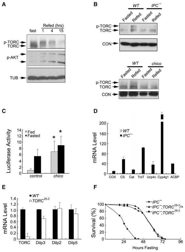
Figure 4. The Insulin Signaling Pathway Regulates TORC Activity in Drosophila.
(A) Immunoblot of phospho-TORC and total TORC protein in fasted or refed flies. Hours after refeeding are indicated. Amounts of phospho-AKT are also shown.
(B) Immunoblots showing amounts of phospho-TORC and total TORC in WT and insulin-producing cell (IPC)-ablated flies (top) and chico mutant flies (bottom) under fasted or refed conditions. Loss of insulin-like peptide (ilp) gene expression in IPC-ablated flies was confirmed by qPCR analysis (data not shown).
(C) Comparison of CRE-luc reporter activity in WT and chico mutant flies under fed or fasted conditions. n = 13 per group; p < 0.05.
(D) qPCR analysis showing relative expression of TORC-regulated genes in WT and IPC-ablated flies.
(E) qPCR analysis of ilp2, ilp3, and ilp5 gene expression in WT and TORC25-3 flies fed ad libitum.
(F) Effect of IPC ablation on starvation sensitivity in TORC25-3 flies.
Data are given as means ± SD.

