
Figure 6. The maturation of M cells in Spib−/− bone marrow chimeric mice.
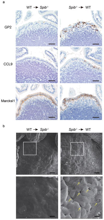
(a) Bone marrow (BM) cells from wild-type (CD45.1) mice were transferred into irradiated Spib−/− (CD45.2) mice (WT → Spib−/−, left column), or vice versa (Spib−/−→ WT, right column). Eight weeks after the transfer, PPs were immunostained with M-cell markers: GP2, CCL9, and Marcksl1, as described in the Experimental Procedures. Scale bar: 50 μm. (b) SEM images of PPs from BM chimeric Spib−/− mice (WT → Spib−/−, left column) and BM chimeric wild-type mice (Spib−/−→ WT, right column) are shown. Lower panels show enlarged images of squares in upper panels. Scale bar = 20 μm (upper), 6 μm (lower). Data are representative of two independent experiments.