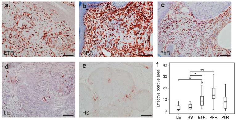
Figure 5. Localization and density of fibroblasts (FBs)/cytes and mesenchymal structures of blood vessels in human skin, as shown by immunohistochemistry and quantitative analysis of stained dermis.
Immunoreactivity for vimentin was observed in erythematous rosacea (ETR, n = 9), papulopustular rosacea(PPR, n = 9), phymatous rosacea (PhR, n = 9), lupus erythematosus (LE; n = 9), and healthy skin (HS; n = 10; bar = 100 μm; a–e). Density of FBs was increased in all subtypes, but significantly so in ETR (× 2.9) and PPR (× 3.94). Skin of lupus patients showed decreased density of FBs/cytes as compared with healthy human skin (*P<0.05; **P<0.01; unfilled circle represents outlier) (f).

