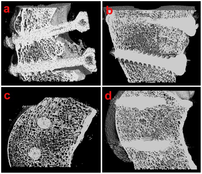
Figure 6. The micro-CT images of screws-bone interface taken at 12 weeks after operation.
(a) Coronal micro-CT image of bioactive screw-bone interface. (b) Coronal micro-CT image of metallic screw-bone interface. (c) Sagittal micro-CT image of the bioactive screw-bone interface. (d) Sagittal micro-CT image of the metallic screw-bone interface.

