
Figure 1.
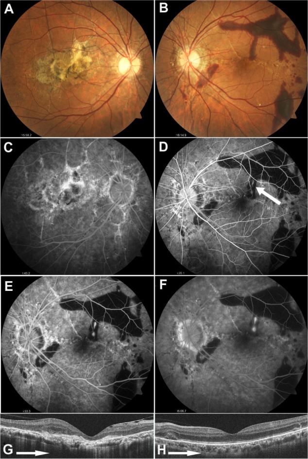
Fundus findings on initial examination.
Notes: (A and B) Fundus photographs: Choroidal atrophy around the optic papillae and angioid streaks radiating from around the optic discs are evident in both eyes. (A) Atrophic changes to the macular region are seen in the right eye, and (B) subretinal hemorrhage within the vascular arcade is apparent in the left; (C–F) Fluorescein angiograms 2 weeks after the onset: (C) Tissue staining and window defect are present in the macular region of the right eye. (D) The early phase image (25 seconds after injection) of the left eye reveals signs of blocking due to subretinal hemorrhage, and hyperfluorescent spots (arrow) due to early leakage superior to the fovea. The images of (E) the middle phase (80 seconds after injection) and (F) the late phase (10 minutes after injection) show increasing hyperfluorescence due to leakage, suggestive of extrafoveal choroidal neovascularization; (G and H) Optical coherence tomography: (G) Thinning of the macula is seen in the right eye, but (H) the left eye shows no obvious abnormality in the neighborhood of the fovea.