
Figure 4.
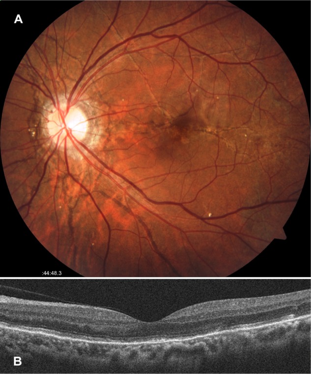
Left fundus findings 2 months after the intravitreal injection of bevacizumab.
Notes: (A) Fundus photograph reveals pronounced regression of choroidal neovascularization (CNV); (B) Optical coherence tomography by horizontal scan shows disappearance of subfoveal CNV.