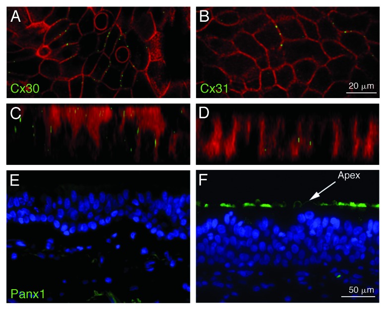
Figure 4. Localization of connexins and Panx1 in airway epithelial cells. These cells express the two connexins, Cx30 and Cx31, which yield the typical punctate staining of gap junctions at the basolateral membranes of contacting cells (A and B). Z-stacks (C and D) do not reveal staining of connexins at the apical membrane. In contrast, staining for Panx1 is restricted to the apical membrane where ATP is released from these cells (E and F). No staining at the basolateral membrane can be detected. (A–D) are from Wiszniewski et al. (2007),34 with permission from Elsevier. (E and F) are adapted from Ransford et al. (2009),24 reprinted with permission of the American Thoracic Society. © Copyright American Thoracic Society.

An official website of the United States government
Here's how you know
Official websites use .gov
A
.gov website belongs to an official
government organization in the United States.
Secure .gov websites use HTTPS
A lock (
) or https:// means you've safely
connected to the .gov website. Share sensitive
information only on official, secure websites.
