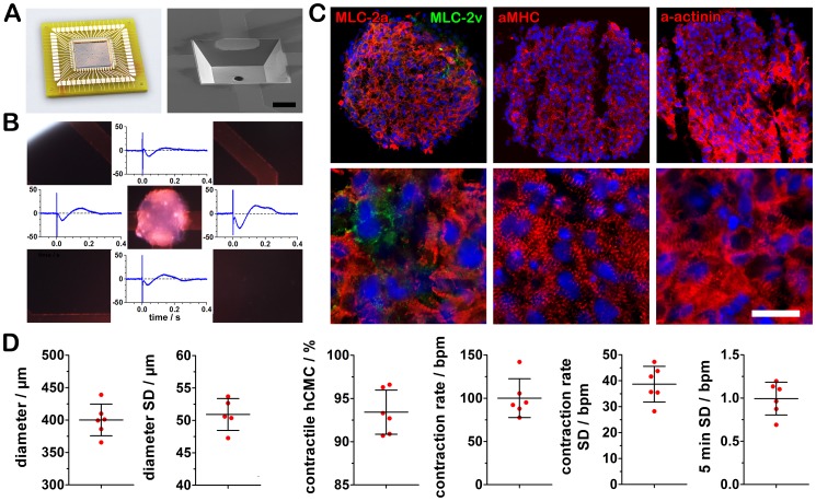
Figure 1. Optimized microcavity array for field potential analysis of highly purified cardiomyocyte clusters.
(A) Bonded silicone-based microcavity array and SEM image of a cavity (400 µm size) with an integrated suction hole for automated positioning. (B) Microcavity array technology enables an instant and detailed field potential recording on four electrodes per cluster without any adhesion requirement of the cardiomyocyte cluster to the electrodes. (C) The hCMC consist of highly enriched cardiomyocytes with atrial (MLC-2a) and to a smaller amount of ventricular cardiomyocyte characteristics (MLC-2v), (bar = 100 µm, zoom: bar = 20 µm). (D) Size and electrophysiological characteristics of six independent differentiation experiments each with 36 hCMC (mean ± s.d.).

