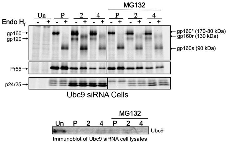
Figure 6. Env and Gag stability in Ubc9 knockdown cells in the presence of proteasome inhibitor MG132.
293T cells were transfected with Ubc9 siRNA and pNL4-3 as in previous experiments. MG132 (10µM) was added to the culture media for 1 hour prior to pulse chase experiments and was maintained throughout the experiment. Cells were pulse (P) labeled with [35S] methionine/cysteine for 1 hour and then chased for 2 and 4 hours. Cell and media associated viral proteins were solublized and immunoprecipitated with pooled AIDS patient sera, split equally, and incubated for 3.5 hours at 37°C in the presence, or absence of Endoglycosidase H f (Endo H f). Samples were separated by SDS PAGE and visualized by phosphorimaging using The Discovery Series Quantity One software. A representative, over-exposed gel is shown so that partially Endo Hf resistant Env can be more easily visualized. Viral proteins and their positions in the gel are labeled on the left. The identity of Endo Hf, untreated viral proteins and their positions in the gel are labeled on the right. Deglycosylated Endo Hf sensitive forms gp160 residing in the ER are labeled as gp160s. Partially deglycosylated, Endo Hf resistant forms of gp160 that have had their glycans modified in the TGN are labeled as gp160r. Forms of gp160 that have undergone glycan modification in the TGN but have not been cleaved are denoted as gp160* in Endo Hf untreated samples.

