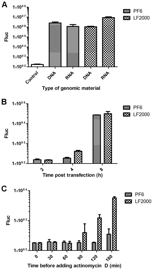
Figure 4. Analysis of Fluc expression in cells transfected with SFV replicon vectors.

(A) BHK-21 cells were transfected with 1 µg of pCMV-SFV(Fluc) 1-EGFP DNA or 1 µg of SFV(Fluc) 1-EGFP RNA using the PF6 or LF2000 reagents. Fluc activities were measured at 14 h post transfection. (B) BHK-21 cells were transfected with 1 µg of pCMV-SFV(Fluc) 1-EGFP DNA using the PF6 or LF2000 reagents. Fluc activities were measured at 2 h, 4 h or 8 h post transfection. (C) BHK-21 cells were transfected with 1 µg of pCMV-SFV(Fluc) 1-EGFP DNA using the PF6 or LF2000 reagents. Actinomycin D (final concentration, 20 µg/ml) was added to the transfection medium immediately (“0” time point) or at time points indicated on the horizontal axes. Fluc activities in transfected cell cultures were measured at 8 h post transfection. In all panels, Fluc activities shown on the vertical axes are normalized to the amount of total protein. Experiments were performed in triplicate, and error bars indicate standard deviations.
