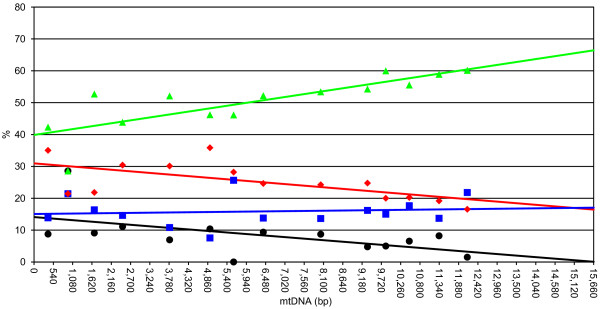
Figure 4.

Location of the origin of replication of the H strand. A (green), C (blue), G (black), and T (red) content at four-fold degenerate codons of PGCs are shown. Percent contents of each PCG are plotted at the midpoint of the ORF using the first nucleotide of cox3 ORF as the starting point. We also included ORF117 in this analysis (see text for further details). Equations are as follows. %A, y = 0.0017 × + 39.88, r2 = 0.64, p < 0.005;%C, y = 0.0001 × 15.07, r2 = 0.01, p = 0.71;%G, y = − 0.0009 × 14.09, r2 = 0.30, p < 0.05;%T, y = − 0.0009 × 30.96, r2 = 0.39, p < 0.05.
