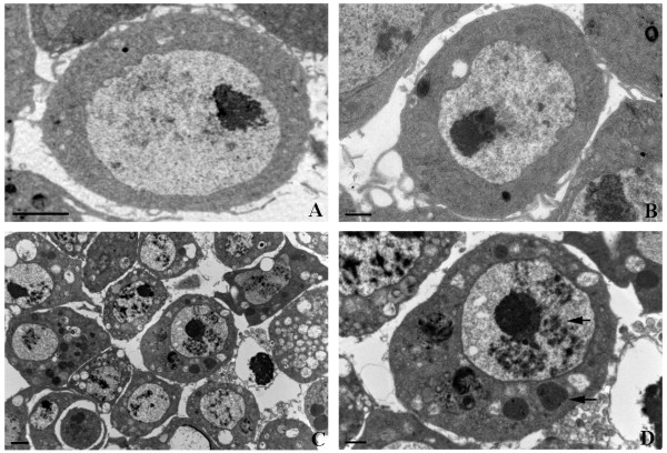
Figure 5.

Transmission electron microscopy (TEM) of DCV-infected S2 cells. (A) Normal S2 cells, Bar=2 μm. (B) DCV-infected S2 cells at one hour post infection, Bar=1 μm. (C) DCV-infected S2 cells at one day post infection, Bar=2 μm. (D) Enlarge of one DCV-infected S2 cell at one day post infection, Bar=1 μm. The arrows indicate the typical morphological changes of S2 cell caused by DCV infection.
