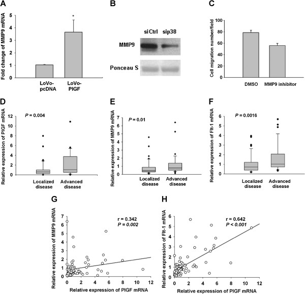
Figure 3.
PlGF induced MMP9 expression via p38 MAPK activation. (A) MMP9 expression was significantly increased in LoVo-PlGF cells compared to LoVo-pcDNA cells as assessed by quantitative PCR. (B) Inhibition of p38 by siRNA (30 pmole/ml) decreased the expression of MMP9 in LoVo-PlGF cells as detected by Western blotting. (C) By using the chemical inhibitor of MMP9 (30 μM), the migration ability was inhibited in LoVo-PlGF cells. In human colorectal cancer tissue, PlGF (D), MMP9 (E) and Flt-1 (F) expression were significantly higher in the advanced than localized disease group. There was a strongly positive correlation between the expression of PlGF and MMP9 (G), as well as PlGF and Flt-1 (H). * indicated as P < 0.05.

