
Figure 5.
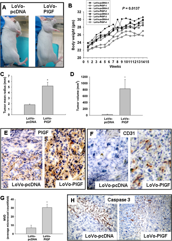
Tumors grew faster and larger in animals receiving LoVo-PlGF xenografts. (A) At the 10th week after injection, tumors were visible in the LoVo-PlGF group but not in the LoVo-pcDNA group. (B) The LoVo-PlGF group lagged behind in body weight gain. Both the tumor radius (C) and tumor volumes (D) were larger in the LoVo-PlGF group than the control group. (E) PlGF expression was significantly higher in the tumor tissue induced by LoVo-PlGF cells. (F) Angiogenesis was also more advanced in the LoVo-PlGF group, with the microvessel density shown in (G). (H) Less apoptosis in LoVo-PlGF group than the control group. * indicated as P < 0.05.