Fig. 4.
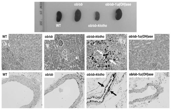
Macroscopic and microscopic changes in the kidney and aorta. Gross morphology of the kidneys from the wild-type (WT), ob/ob, ob/ob-klotho−/− and ob/ob-1α(OH)ase−/− mice. Compared to the ob/ob and ob/ob-1α(OH)ase−/− mice, the kidney sizes were smaller in the hyperphosphatemic ob/ob-klotho−/− mice. All animals were age-matched (11 weeks) (upper panel). Histological sections of kidneys were prepared from WT, ob/ob, ob/ob-klotho−/− and ob/ob-1α(OH)ase−/− mice (middle panel). Inducing phosphate toxicity in the ob/ob mice resulted in extensive calcifications (dark black staining, arrows) in the kidneys of the ob/ob-klotho−/− mice. In contrast, reducing the serum phosphate levels in ob/ob mice did not show any calcification in the ob/ob-1α(OH)ase−/− mice (20× magnification). Aortic sections were prepared from all mice (lower panel). Inducing phosphate toxicity in the ob/ob mice resulted in extensive aortic calcifications (dark black staining, arrows) compared to the ob/ob-klotho−/− mice. In contrast, reducing serum phosphate levels in the ob/ob mice did not lead to any calcification (von Kossa staining; 20× magnification).

