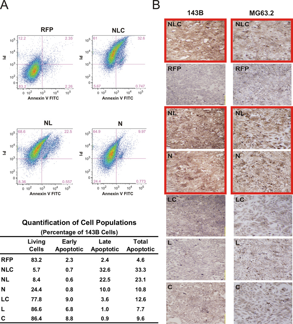
Figure 3. IGFBP5 deletional mutants’ effects on apoptosis.
A. Apoptosis by flow cytometry: 143B cells were transduced with the indicated constructs. Cells were stained with propidium iodide & AnnexinV and assessed for apoptosis by flow cytometry. The cells were clustered according to those undergoing early and late phases of apoptosis. Distributions of the populations for RFP, NLC, NL, and N constructs are shown in the top panel. Quantification of the alive, early apoptotic and late apoptotic populations for all constructs is shown in the bottom panel. B. Caspase 3 staining: 143B and MG63.2 cells were transduced with the indicated adenoviruses, and immunohistochemistry staining for cleaved caspase 3 performed as an indicator of apoptosis was performed. Note the increased staining in the N-terminal domain-containing groups (outlined red).

