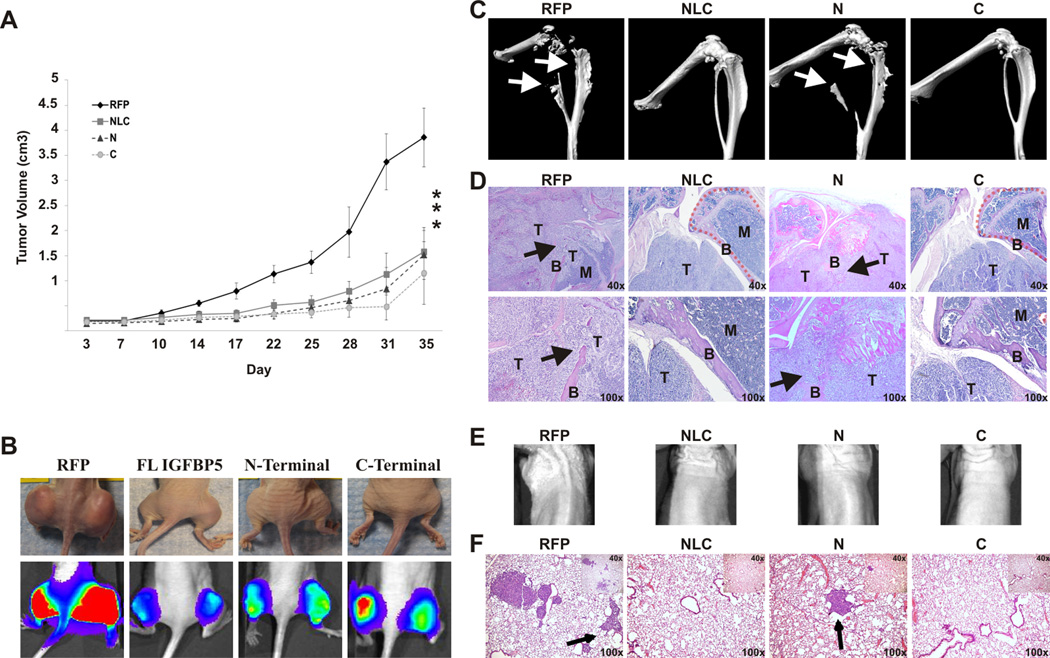
Figure 6. In vivo tumor and metastases.
A. Primary tumor growth of 143B cells expressing control RFP, the full-length NLC, N-terminal or C-terminal constructs: At 36 hours, cells were harvested and injected subperiosteally into the bilateral proximal posterior tibiae of athymic nude mice. Tumors were measured and subjected to live bioluminescent imaging every 3–4 days. The tumor volume and doubling time were calculated. At five weeks, animals were sacrificed and primary tumors and lungs were harvested for evaluation. The asterisk (*) indicates a significant difference in doubling time compared to the RFP control (p < 0.01).
B. Representative photographic and bioluminescent images of the primary tumors of mice for each of the indicated groups: Bioluminescent imaging and quantitative analysis was performed every 3–4 days. The photographs and bioluminescent images seen are at four days prior to tumor harvesting. C. MicroCT of the primary tumors: Note the bone destruction of the tibia and fibula (arrows) in the RFP and N-terminal constructs. Conversely, the full-length NLC and C-terminal constructs had normal appearing tibias and fibulas. D. Hematoxylin and eosin staining of the primary tumors: Note that the cortex of the tibia is intact in the full-length NLC and C-terminal groups (dashed lines), whereas that bone structure is lost in the RFP and N-terminal groups due to tumor invasion. In the RFP and N-terminal groups, the bone cortex (B) is destroyed by the tumor (T), and the bone marrow space (M) is being replaced by tumor. E. Representative bioluminescent images of the thorax of the mice four days prior to sacrifice. F. Lung metastases: Representative hematoxylin and eosin stained sections of the lungs of animals from the indicated groups are shown. Note the metastases seen in the RFP and the N-terminal groups (arrow), whereas the full-length NLC and C-terminal groups show minimal metastases.

