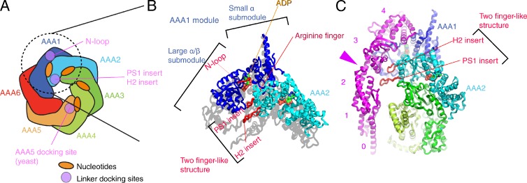
Figure 4.
Structures around the first ATP binding site. (A) Schematic domain structure of the head domain. Regions contacting the linker domain are colored purple. (B) AAA submodules surrounding the first nucleotide-binding pocket (PDB accession no. 3VKG, chain A). The linker is connected to AAA1 domain by the “N-loop.” To highlight that the two finger-like structures are protruding, the shadow of the atomic structure has been cast on the plane parallel to the head domain. (C) Interaction between the linker and the two finger-like structures. The pink arrowhead points to the hinge-like structure of the linker. The pink numbers indicates the subdomain of the linker.

