Figure 4.
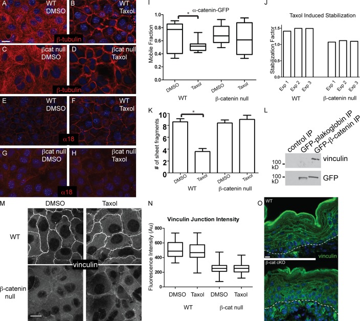
β-Catenin–null cells do not undergo microtubule-dependent adherens junction engagement and epithelial sheet strengthening. (A–D) WT or β-catenin (βcat)–null keratinocytes were treated with DMSO or 10 µM taxol for 1 h before fixation and staining with anti–β-tubulin antibodies. (E–H) WT or β-catenin–null cells (control and taxol treated) were stained with α18 antibodies, which recognize a tension-sensitive epitope of α-catenin. (I) FRAP was performed on WT and β-catenin–null cells expressing α-catenin–GFP. Mobile fraction was determined, and a representative experiment is shown. A significant decrease in mobile fraction was detected only in WT cells treated with taxol, as compared with control (*, P < 0.05). Horizontal lines are the median. Boxes are 25–75%, and whiskers are 10–90%. (J) We determined the stabilization factor (median mobile fraction of control cells/median mobile fraction of taxol-treated cells) for three independent experiments for both WT and β-catenin–null cells. Exp, experiment. (K) Confluent monolayers of WT or β-catenin–null cells were treated with DMSO or 10 µM taxol for 1 h before dispase treatment to release the monolayer from the underlying substrate. After mechanical disruption by pipetting, the number of cell sheet fragments was counted. Larger numbers indicate more fragile sheets, whereas fewer is indicative of increased mechanical integrity. Error bars are standard deviations. (L) Immunoprecipitates (IP) of GFP-tagged plakoglobin or β-catenin were probed with antibodies again vinculin and GFP. (M) WT and β-catenin–null cells were either DMSO or taxol treated for 1 h before fixation and staining with anti-vinculin antibodies. (N) Quantitation of vinculin cortical intensity under the indicated conditions. Horizontal lines are the median. Boxes are 25–75%, and whiskers are 10–90%. Au, arbitrary units. (O) Vinculin localization in WT and β-catenin–null epidermis. The dotted lines indicate the basement membrane. Bars, 10 µm.

