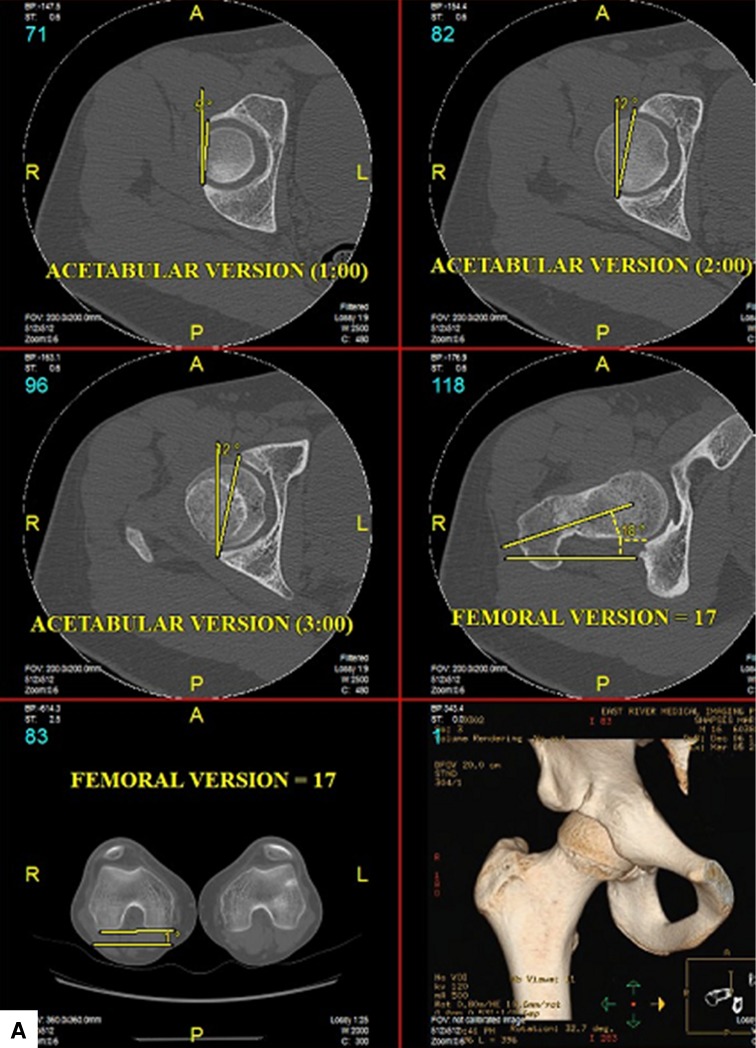
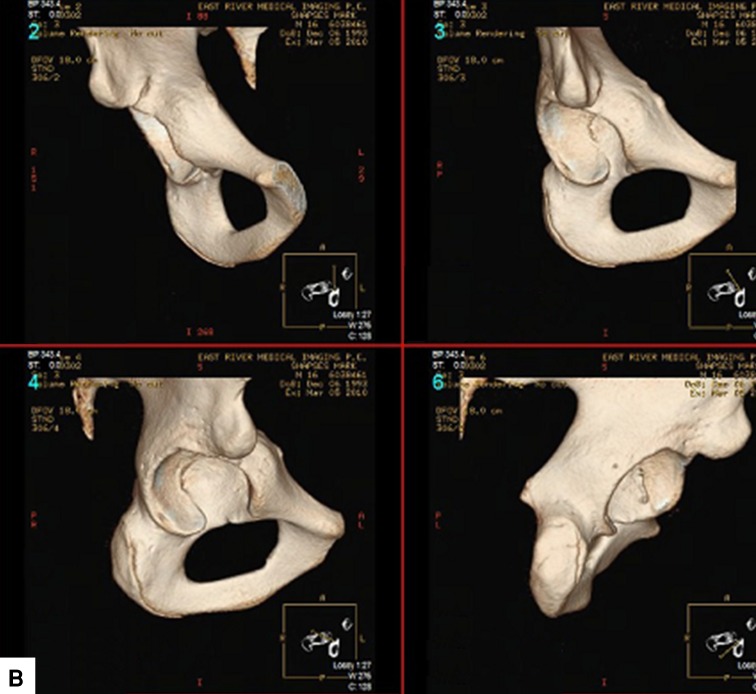
Fig. 5A–B.
(A) Axial CT images of the right hip demonstrate anteversion of the acetabulum at the 12-, 1-, and 2-o’clock locations despite the radiographic appearance of a crossover sign. Femoral version is also assessed at 17° as referenced from the posterior condylar axis of the distal femur. (B) Type III AIIS morphology is shown in AP view (upper left), one-rotation or head-on view (upper right), two-rotation view (lower left), and four-rotation or ischium view (lower right). Part of the AIIS crosses caudad to the horizontal line and also caudad to the anterior superior acetabular rim.


