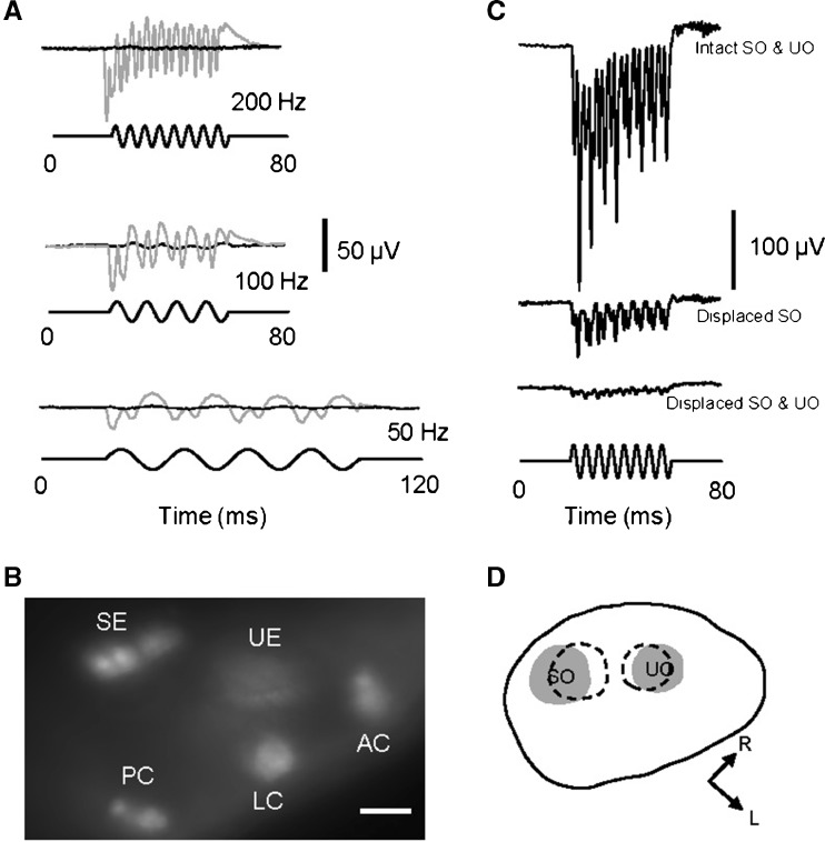
FIG. 3.
A Microphonic responses before (gray traces) and after (black traces) an injection of a fluorescent dye, FM1-43X, into the otic vesicle of a 3-dpf zebrafish in response to 50-Hz (bottom), 100-Hz (middle), and 200-Hz (top) stimuli at 5.8-μm displacement. B Fluorescent image of saccular and utricular epithelia (SE and UE) and anterior, lateral, and posterior cristae (AC, LC, and PC) that were labeled with FM1-43X after the injection. The image was taken from the top view of the zebrafish. Scale bar = 20 μm. C Microphonic responses from a larva with intact saccular and utricular otoliths (top row of response), the displacement of the saccular otolith alone (middle row of response), and the displacement of both otoliths (bottom row of response). Stimuli: 200 Hz at 5.8-μm displacement. D Drawing of the otic vesicle of the larva shown in C, illustrating the original SO and UO positions (gray areas) and displaced positions (dashed areas). L lateral of the larva, R rostral.

