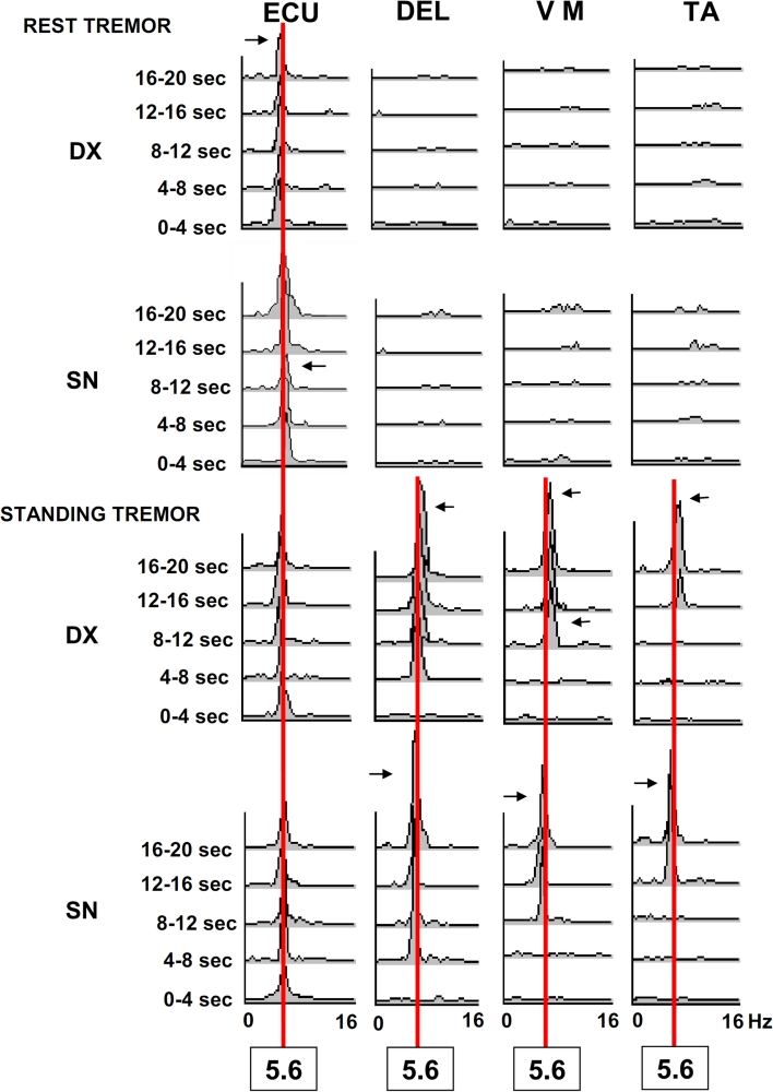
Fig. 3.
Frequency analysis and power spectra of tremor at rest and during standing with wing beating arm posture in a patient affected by DLB. EXTENSOR CARPI ULNARIS = ECU; DELTOID = DEL; VASTUS MEDIALIS = VM; TIBIALIS ANTERIOR = TA Horizontal scales represent frequencies in Hz, (0–16 Hz) amplitude of random noise frequencies are 0.2–0.6 μV2. A vertical red line connects 5.6 Hz frequencies, which is the specific tremor frequency in this patient. Horizontal arrows point to minor frequency variations (range 0.5–01 Hz) of the peak amplitude. Notice overflow during standing posture for the same districts involved by the rest tremor to other body districts

