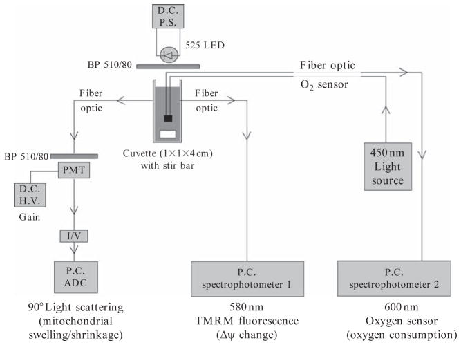
Figure 14.4.
Configuration of mitochondrial assay system. Mitochondria preparation is suspended in stirred cuvette. Excitation source is light emitting diode (LED) and BP 510/80 Fluorescence filter. Mitochondrial swelling isdetected by 90 degree scattered light at photomultipler tube (PMT).TMRM fluorescence is detected by Spectrophotometer1, and oxygen is detected using a fiber–optic sensor inside of the mitochondrial solution by Spectrophotometer 2. Signals are logged by analog to digital converter (ADC) in a PC.

