Figure 3.
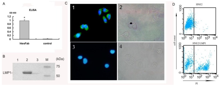
Characterization of Fab binding with LMP1-TES1 domain. (A) ELISA showed that htesFab bound LMP1-TES domain in native confirmation (p < 0.05); (B) Immunoprecipitation analysis for the detection of LMP1 protein. LMP1 was 53 kDa; Line 1: HNE2 cells; line 2: HNE2-LMP1 cells; line 3: unrelated Fab fragment (C); Immunofluorescence analysis showed that htesFab labeled LMP1 in the intracellular and plasma membranes in HNE2-LMP1 cells (green), cell nuclei were stained with DAPI (blue) (×200, Olympus digital camera); (D) FACS analysis showed the binding of htesFab to HNE2-LMP1(40.35%) and HNE2 cells(4.15%) (blue dots). Background staining was obtained by PBS.

